Comprehensive State-of-the-Art Eye Examinations
Our comprehensive eye examinations evaluate your eyes, both inside and out, for any potential disorders or diseases. Your visual skills and abilities are carefully evaluated and appropriate treatment is prescribed – whether it’s medication, lenses or low vision rehabilitation.
Wavefront optimized refraction represents the state of the art in refraction-an accurate assessment of the patients total visual system, in a fraction of the time.
 Most practices still perform what is called a manual refraction. You know, the part of the examination when we ask you, “Which one is better, one or two?” Although this subjective comparison is still critically important, the manner in which we perform this test is outdated and some patients dread this part of a comprehensive eye examination.
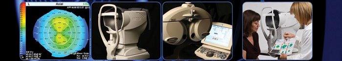
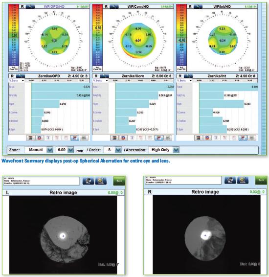
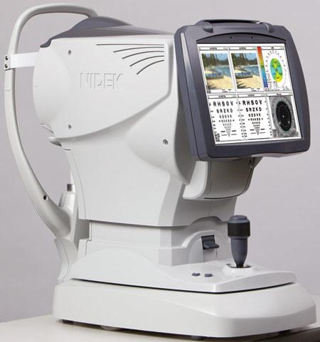
Our office has taken the next step. In fact, we are the first in western Lancaster County to take this step. We perform a wavefront optimized refraction as a part of our comprehensive examination. Scientist have discovered that there is much more to measuring your overall vision. In fact, we now know that the manner in which light refracts into the eye and the way we process this information visually is unique to every individual-much like a finger print. We can now measure these fine differences with the Marco OPD Scan III. This instrument houses six different instruments in one allowing us to take measures with great efficiency and accuracy.
After the OPD Scan is performed, we compare all data and send a summary of this information to the Marco TRS-5100 automatic refractor.
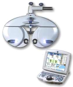
The TRS 5100 replaces the standard refractor designed in 1928 and which most offices use. It allows us to control the entire refraction process from a keypad.
 Prescription lens choices move quickly, more accurately and efficiently providing less discomfort to you. Because the TRS 5100 is completely programmable, all the lenses are moved at the touch of a button, taking us to each new refraction step in more efficient and accurate way. The TRS 5100 Refractor is extremely thorough and it decreases the time you need to spend making choices.
For most of the last century, testing vision meant giving the patient the choice between two lenses, “which is better one or two”. In some cases, we can now show you 2 images at the same time to help fine tune your astigmatism to within an unprecedented 1 degree accuracy.
With the TRS-5100 we are able to instantly show you the new prescription compared to the old prescription with a touch of a button so you can make a better decision if updating your glasses will improve your vision. Our patients love the accuracy, speed, and the precision that the TRS 5100 technology provides.
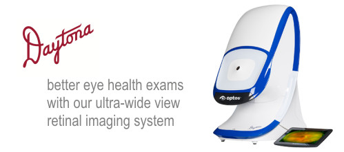
Optomap Pan-retinal Ophthalmoscopy
The Optomap Daytona uses a low-power scanning laser to obtain an ultra-widefield digital image of the retina. With an Optomap image, up to 80% of the retina can be viewed without dilating drops. This allows Dr. Rebman and Dr. Zechman to confirm your eye is healthy or better detect diseases like macular degeneration, diabetic retinopathy, glaucoma, melanoma, etc. The Optomap creates a permanent record for your file, which allows us to view your images each year to look for changes.
Optical Coherence Tomography (OCT)
This is a non-invasive imaging test that uses light waves to take cross-section pictures of your retina, the light-sensing tissue lining the back of the eye. OCT is a sophisticated scanning system that produces highly detailed images of the retina and optic nerve. It is often likened to an MRI or x-ray of the eye. This scan allows Dr. Rebman and Dr. Zechman to see detailed images of the retina (the inner most layer of the interior eye) and optic nerve, enabling them to accurately detect and monitor changes to the retina and optic nerve. OCT is especially useful in monitoring and treating macular degeneration and glaucoma.
Corneal Topographer
The corneal topographer measures the curvature and shape of the cornea found on the front surface of the eye. The data obtained creates a topographical map of the corneal surface. This test is helpful in diagnosing diseases or degenerations of the cornea like keratoconus. It may also be used to successfully fit contact lenses, monitor ortho-keratology, and determine if an individual is a good candidate for refractive surgery like LASIK.